Patient care services are open to the public. Appointments can be made by calling (231) 591-2020.
1124 S. State Street, MCO 101A
Big Rapids MI 49307
(231) 591-2020
(231) 591-3991 (fax)
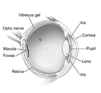
A transparent liquid in the front 1/3 of the eye, filling the area between the cornea and lens.
The transparent front window of the eye. The cornea provides most of the power necessary for focusing light onto the retina.
A transparent, biconvex structure located behind the iris of the eye, it is responsible for additional power for focusing light onto the retina Photo courtesy of Dr. Mark Swan
A small area on the retina that is used for central, detailed vision.
Structure at the back of the eye responsible for carrying nerve impulses from the retina to different areas of the brain.
The black circular opening seen in the eye. The pupil changes it's diameter in order to control the amount of light entering the eye.
A transparent gel located in the back 2/3 of the eye, filling in the area between the lens and the retina.

Photo courtesy of National Eye Institute, National Institutes of Health
A vision condition that occurs when the front surface of your eye, the cornea, is slightly irregular in shape. This irregular shape prevents light from focusing properly on the retina. Your vision may be blurred at all distances. People with astigmatism may experience headaches, eye strain, fatigue or blurred vision at certain distances. Most people have some degree of astigmatism.
A bifocal is a lens that has two focusing sections. The top part of the bifocal is usually used for distant/far off vision and the bottom portion is used for reading/near vision.
A chronic or long term inflammation of the eyelids and eyelashes. It affects people of all ages. Among the most common causes of blepharitis are poor eyelid hygiene; excessive oil produced by the glands in the eyelid; a bacterial infection (often staphylococcal); or an allergic reaction.
The lens of an eye is normally clear. If the lens becomes cloudy or is opacified it is called a cataract. Cataracts may be present at or shortly after birth in which case they are called congenital cataracts. Adult cataracts develop with advancing age, tend to run in families, and the appearance may be accelerated by environmental factors.
The ability to distinguish some colors and shades is less than normal. It occurs when the color-sensitive cells in the retina do not properly pick up or send the proper color signals to the brain. About eight percent of men and one percent of women are color deficient. This can mean certain shades of colors are not distinguishable or, in very rare cases, color deficiency exists to an extent that no colors can be detected, only shades of black, white and gray.
A decline in either the tear quality or quantity that bathe the outside area of the cornea. This may lead to symptoms such as: tearing, burning, irritation, or a sandy feeling.
A condition of increased fluid pressure inside the eye (intraocular pressure). Increased pressure occurs when the aqueous humor, which is produced continuously, does not drain properly. The pressure pushes on the retina, reducing the blood supply to the nerves of the retina causing them to die. As the optic nerve deteriorates, blind spots and vision changes develop. Glaucoma is a leading cause of blindness, but the chance can be reduced if caught early and controlled by medication.
Sometimes referred to as farsightedness. Hyperopia is a vision condition in which distant objects are usually seen clearly, but close ones do not come into proper focus. Farsightedness occurs if your eyeball is too short or the cornea has too little power, so light entering your eye is focused behind the retina, instead of directly on the retina, causing blurred vision.
May be recommended by your optometrist to treat conditions such as blepharitis. There are commercially prepared lid scrubs, or your doctor may recommend gently washing your lashes with warm water and baby shampoo.
Sometimes referred to as nearsightedness. Myopia is a vision condition in which near objects are seen clearly, but distant objects do not come into proper focus. Nearsightedness occurs if your eyeball is too long or the cornea is too powerful, so the light entering your eye is focused before the retina, instead of directly onto the retina, causing blurred vision.
An ophthalmologist is a physician (MD or DO) who specializes in eye health and eye disease. Ophthalmologists attend four years of pre-medical college, and then attend four years of medical school. After medical school, an ophthalmologist attends a residency in ophthalmology where they learn about diseases and surgeries of the eye.
An optician is a person who is qualified to fill spectacle lens prescriptions. Opticians are trained to fabricate spectacles, verify prescriptions, mount lenses into frames, adjust glasses, and dispense glasses or contact lenses. In addition, opticians determine the type of lenses best suited for the patient. Opticians attend a two-year associate degree program in opticianry or they may receive their education through on the job training.
An optometrist is a doctor of optometry (OD). Optometrists attend four years of pre-health profession college, and then attend four years of optometry college. After optometry school, some optometrists attend a residency program which can specializes in contact lens, ocular disease, or other areas. Optometrists provide primary eyecare services such as: 1) comprehensive eye and vision evaluations; 2) contact lens evaluations, 3) low vision rehabilitation; 4) diagnosis, treatment and management of eye diseases and vision disorders; 5) binocular vision analysis; 6) vision therapy; 7) medical health anomaly detection; 8) certain surgical procedures; 9) patient consultation regarding visual needs and surgical alternatives; and 10) spectacles and contact lenses prescriptions.
Small, semi-transparent or cloudy specks or particles within the vitreous. They appear as specks of various shapes and sizes, threadlike strands or cobwebs. Since they are within your eyes, they move as your eyes move and seem to dart away when you try to look at them directly. Spots are often caused by small flecks of protein or other particles trapped during the formation of your eyes before birth. They can also result from deterioration of the vitreous due to aging or from certain eye diseases or injuries. Most spots are not harmful and rarely limit vision. But, spots can be indications of more serious problems, and you should see your optometrist for a comprehensive examination when you notice sudden changes or see increases in them.
A trifocal is a lens that has three focusing sections. The top part of the bifocal is usually used for distant/far off vision. The middle section, known as the intermediate, is used for arms distance length viewing such as viewing dashboards on cars or computer screens. The bottom portion is used for reading/near vision.
20/20 vision is a term used to express normal visual acuity (the clarity or sharpness of vision) measured at a distance of 20 feet. If you have 20/20 vision, you can see clearly at 20 feet what should normally be seen at that distance. If you have 20/100 vision, it means that you must be as close as20 feet to see what a person with normal vision can see at 100 feet. 20/20 does not necessarily mean perfect vision. 20/20 vision only indicates the sharpness or clarity of vision at a distance. There are other important vision skills, including peripheral awareness or side vision, eye coordination, depth perception, focusing ability and color vision that contribute to your overall visual ability.
Clinic Referral Forms-(Professional)
HIPAA privacy forms and information
Patient care services are open to the public. Appointments can be made by calling (231) 591-2020.
1124 S. State Street, MCO 101A
Big Rapids MI 49307
(231) 591-2020
(231) 591-3991 (fax)

